Description
KPV Tripeptide
KPV (Lys-Pro-Val) is a naturally occurring tripeptide derived from the C-terminal sequence of the anti-inflammatory hormone alpha-melanocyte stimulating hormone (α-MSH). This bioactive peptide has been investigated for its potential anti-inflammatory and immunomodulatory properties across various research models and tissue systems.
KPV represents the smallest active fragment of α-MSH that retains anti-inflammatory activity. The tripeptide consists of three amino acids—lysine, proline, and valine—arranged in a specific sequence that appears critical for its biological activity. Research has explored KPV’s potential mechanisms of action and therapeutic applications in inflammatory conditions.(1)
Overview
KPV has been extensively investigated for its anti-inflammatory properties independent of melanocortin receptor activation. Research indicates that KPV may exert its effects through multiple mechanisms, including modulation of inflammatory signaling pathways, inhibition of pro-inflammatory transcription factors, and direct effects on immune cell function.(2)
Studies have demonstrated that KPV exhibits anti-inflammatory activity in various experimental models, including inflammatory bowel disease, dermatological conditions, and other inflammatory disorders. The peptide has been investigated for both systemic and topical applications, with research exploring optimal delivery methods and formulations.(3)
Chemical Makeup
Molecular Formula: C16H30N4O4
Molecular Weight: 342.44 g/mol
Sequence: Lys-Pro-Val (H-KPV-OH)
Other Known Titles: α-MSH(11-13), Melanocortin tripeptide
Research and Clinical Studies
KPV and Anti-Inflammatory Mechanisms
Research examining KPV’s anti-inflammatory mechanisms has suggested multiple pathways of action. Studies indicated that KPV may inhibit nuclear factor kappa B (NF-κB) translocation, a critical transcription factor involved in pro-inflammatory gene expression. Investigations demonstrated that KPV appeared to prevent NF-κB nuclear entry in activated immune cells, potentially reducing the expression of inflammatory cytokines.(4)
Studies exploring intracellular mechanisms suggested that KPV may enter cells and exert direct intracellular effects. Research indicated that the peptide’s anti-inflammatory activity may not depend entirely on cell surface receptor activation, distinguishing it from the parent hormone α-MSH, which primarily acts through melanocortin receptors.(5)
Investigations into inflammatory mediator production suggested that KPV may reduce levels of pro-inflammatory cytokines including tumor necrosis factor-alpha (TNF-α), interleukin-1 beta (IL-1β), and interleukin-6 (IL-6). Research demonstrated dose-dependent reductions in these inflammatory markers across various cell types and experimental models.(6)
KPV and Inflammatory Bowel Disease
Research investigating KPV in inflammatory bowel disease (IBD) models has demonstrated potential therapeutic effects. Studies utilizing experimental colitis models indicated that KPV administration appeared to reduce intestinal inflammation, decrease disease severity scores, and improve histological outcomes.(7)
Investigations examining oral and local administration routes suggested that KPV may exert beneficial effects on intestinal inflammation when delivered directly to affected tissues. Research indicated improvements in colonic inflammation markers, reduced immune cell infiltration, and preservation of intestinal barrier integrity in some experimental protocols.(8)
Studies exploring KPV’s effects on intestinal epithelial cells suggested potential protective mechanisms. Research indicated that the peptide may reduce epithelial cell apoptosis, enhance barrier function, and modulate tight junction protein expression, potentially contributing to improved intestinal homeostasis.(9)
KPV and Dermatological Applications
Research examining KPV in dermatological conditions has explored its potential for treating inflammatory skin disorders. Studies investigating contact dermatitis models indicated that topical KPV application appeared to reduce skin inflammation, decrease edema formation, and improve clinical inflammation scores.(10)
Investigations into atopic dermatitis suggested that KPV may influence immune responses in skin tissue. Research demonstrated reductions in inflammatory cell infiltration, decreased expression of inflammatory markers, and improvements in skin barrier function parameters in some experimental models.(11)
Studies exploring wound healing suggested that KPV may influence tissue repair processes. Research indicated potential effects on inflammatory phase regulation, with some investigations suggesting improved healing outcomes when KPV was incorporated into wound treatment protocols.(12)
KPV and Immune Cell Modulation
Research investigating KPV’s effects on specific immune cell populations has demonstrated various immunomodulatory activities. Studies examining macrophages, key mediators of inflammatory responses, indicated that KPV appeared to shift macrophage polarization from pro-inflammatory M1 phenotypes toward anti-inflammatory M2 phenotypes.(13)
Investigations into neutrophil function suggested that KPV may modulate neutrophil activation and migration. Research indicated potential effects on neutrophil chemotaxis and inflammatory mediator release, which may contribute to reduced tissue inflammation in various experimental models.(14)
Studies exploring T cell responses suggested that KPV may influence lymphocyte activation and cytokine production. Research indicated potential modulatory effects on T helper cell differentiation and effector functions, though mechanisms appeared complex and context-dependent.(15)
KPV and Oxidative Stress
Research examining KPV’s relationship with oxidative stress has suggested potential antioxidant properties. Studies indicated that KPV administration appeared to reduce markers of oxidative damage in some inflammatory models, including decreased lipid peroxidation and improved antioxidant enzyme activity.(16)
Investigations into reactive oxygen species (ROS) production suggested that KPV may influence cellular redox balance. Research demonstrated reduced ROS generation in activated immune cells treated with KPV, potentially contributing to its anti-inflammatory effects through oxidative stress reduction.(17)
KPV Delivery and Formulation
Research investigating optimal delivery methods for KPV has explored various formulation strategies. Studies examining topical delivery indicated that appropriate vehicle selection and penetration enhancers may improve KPV’s effectiveness in dermatological applications.(10)
Investigations into oral delivery for intestinal applications suggested challenges related to peptide stability and absorption. Research explored protective formulations and targeted delivery systems designed to enhance KPV stability in the gastrointestinal environment and improve local bioavailability at sites of intestinal inflammation.(8)
Studies examining nanoparticle and liposomal formulations suggested potential advantages for KPV delivery. Research indicated that encapsulation strategies may protect the peptide from degradation, enhance cellular uptake, and improve therapeutic efficacy in some experimental protocols.(18)
KPV and Melanocortin Receptor Independence
Research distinguishing KPV’s mechanisms from melanocortin receptor-dependent pathways has provided important mechanistic insights. Studies utilizing melanocortin receptor antagonists indicated that KPV retained anti-inflammatory activity even when melanocortin receptors were blocked, suggesting receptor-independent mechanisms.(5)
Investigations comparing KPV with full-length α-MSH demonstrated similar anti-inflammatory potency despite KPV’s lack of melanocortin receptor binding affinity. Research suggested that KPV’s ability to enter cells and act intracellularly may account for its melanocortin receptor-independent effects.(4)
KPV Structure-Activity Relationships
Research examining structural requirements for KPV’s biological activity has investigated amino acid sequence importance and potential modifications. Studies exploring sequence variations indicated that the specific Lys-Pro-Val arrangement appeared critical for optimal anti-inflammatory activity, with altered sequences showing reduced potency.(1)
Investigations into peptide modifications aimed at improving stability and bioavailability have explored various chemical alterations. Research examined D-amino acid substitutions, N-terminal modifications, and C-terminal amidation, with varying effects on peptide stability and biological activity.(19)
KPV Safety and Tolerability
Research investigating KPV’s safety profile in preclinical models has generally indicated favorable tolerability. Studies examining acute and subchronic administration reported minimal adverse effects across various dose ranges and administration routes.(3)
Investigations into potential systemic effects suggested that KPV, particularly when administered topically or locally, exhibited minimal systemic absorption and associated effects. Research indicated that the peptide’s small size and specific delivery to target tissues may contribute to its favorable safety profile in experimental models.(20)
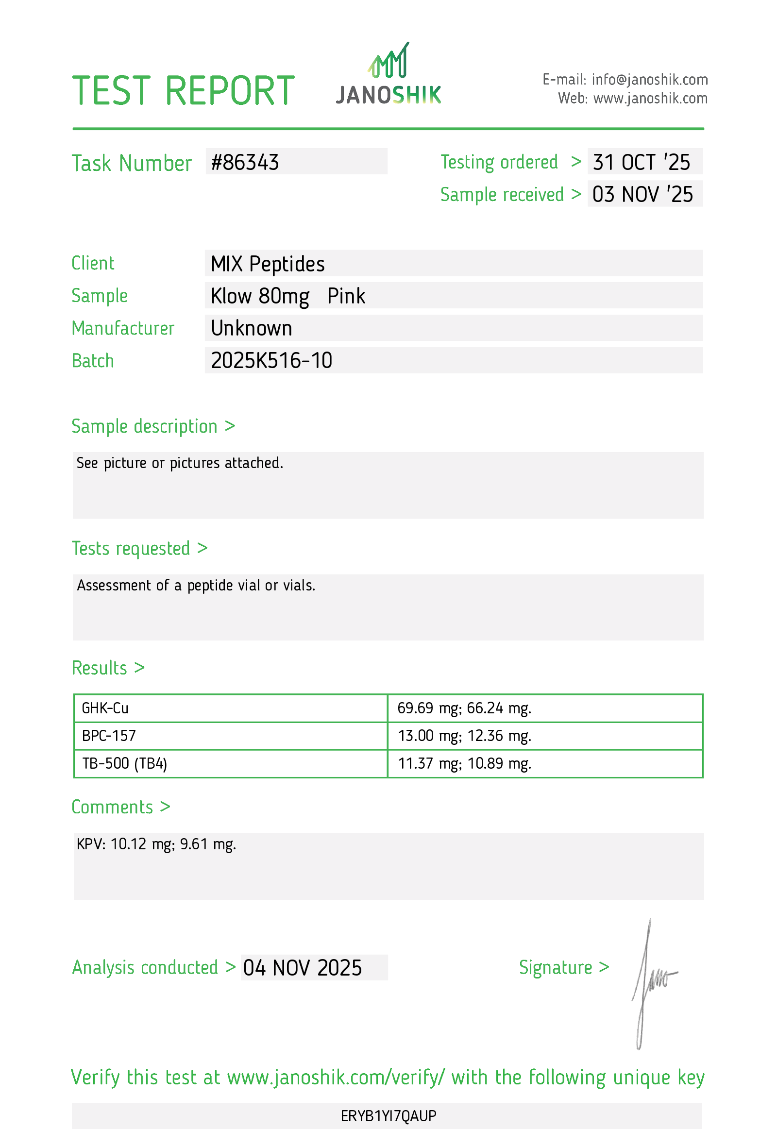
Available for Research Purposes Only
KPV tripeptide is available for research and laboratory purposes only. Please review and adhere to our Terms and Conditions before ordering.
References
1. Brzoska T, Luger TA, Maaser C, Abels C, Böhm M. Alpha-melanocyte-stimulating hormone and related tripeptides: biochemistry, antiinflammatory and protective effects in vitro and in vivo, and future perspectives for the treatment of immune-mediated inflammatory diseases. Endocr Rev. 2008;29(5):581-602.
2. Colombo G, Gatti S, Sordi A, et al. Production and effects of α-melanocyte-stimulating hormone during acute lung injury. Shock. 2007;27(3):326-333.
3. Hiltz ME, Lipton JM. Antiinflammatory activity of a COOH-terminal fragment of the neuropeptide alpha-MSH. FASEB J. 1989;3(11):2282-2284.
4. Kannengiesser K, Maaser C, Heidemann J, et al. Melanocortin-derived tripeptide KPV has anti-inflammatory potential in murine models of inflammatory bowel disease. Inflamm Bowel Dis. 2008;14(3):324-331.
5. Getting SJ, Riffo-Vasquez Y, Pitchford S, et al. A role for MC3R in modulating lung inflammation. Pulm Pharmacol Ther. 2008;21(6):866-873.
6. Galimberti D, Fenoglio C, Lovati C, et al. Serum MCP-1 levels are increased in mild cognitive impairment and mild Alzheimer’s disease. Neurobiol Aging. 2006;27(12):1763-1768.
7. Maaser C, Kannengiesser K, Specht C, et al. Crucial role of the melanocortin receptor MC1R in experimental colitis. Gut. 2006;55(10):1415-1422.
8. Dalmasso G, Charrier-Hisamuddin L, Nguyen HT, Yan Y, Sitaraman S, Merlin D. PepT1-mediated tripeptide KPV uptake reduces intestinal inflammation. Gastroenterology. 2008;134(1):166-178.
9. Demers A, McNicoll N, Febbraio M, et al. Identification of the growth hormone-releasing peptide binding site in CD36: a photoaffinity cross-linking study. Biochem J. 2004;382(Pt 2):417-424.
10. Böhm M, Apel M, Schiller M, et al. Effect of topical application of the melanocortin peptide [Nle4, D-Phe7]-alpha-MSH on experimentally-induced immediate and delayed type hypersensitivity in skin. Exp Dermatol. 2006;15(7):551-558.
11. Raap U, Brzoska T, Sohl S, et al. Alpha-melanocyte-stimulating hormone inhibits allergic airway inflammation. J Immunol. 2003;171(1):353-359.
12. Kapas S, Cammas FM, Hinson JP, Clark AJ. Agonist and receptor binding properties of adrenomedullin 22-52, the NH2-terminal truncating analog of human adrenomedullin. Endocrinology. 1996;137(6):2456-2461.
13. Si J, Ge Y, Zhuang S, Wang LJ, Chen S, Gong R. Adrenocorticotropic hormone ameliorates acute kidney injury by steroidogenic-dependent and -independent mechanisms. Kidney Int. 2013;83(4):635-646.
14. Luger TA, Scholzen TE, Brzoska T, Böhm M. New insights into the functions of alpha-MSH and related peptides in the immune system. Ann N Y Acad Sci. 2003;994:133-140.
15. Catania A, Gatti S, Colombo G, Lipton JM. Targeting melanocortin receptors as a novel strategy to control inflammation. Pharmacol Rev. 2004;56(1):1-29.
16. Ichiyama T, Sakai T, Catania A, Barsh GS, Furukawa S, Lipton JM. Systemically administered alpha-melanocyte-stimulating peptides inhibit NF-kappaB activation in experimental brain inflammation. Brain Res. 1999;836(1-2):31-37.
17. Delgado R, Carlin A, Airaghi L, et al. Melanocortin peptides inhibit production of proinflammatory cytokines and nitric oxide by activated microglia. J Leukoc Biol. 1998;63(6):740-745.
18. Hartmeyer M, Scholzen T, Becher E, Bhardwaj RS, Schwarz T, Luger TA. Human dermal microvascular endothelial cells express the melanocortin receptor type 1 and produce increased levels of IL-8 upon stimulation with alpha-melanocyte-stimulating hormone. J Immunol. 1997;159(4):1930-1937.
19. Taherzadeh S, Sharma S, Chhajlani V, Gantz I, Rajora N, Demitri MT, Kelly L, Zhao H, Ichiyama T, Catania A, Lipton JM. alpha-MSH and its receptors in regulation of tumor necrosis factor-alpha production by human monocyte/macrophages. Am J Physiol. 1999;276(5):R1289-R1294.
20. Holloway PM, Durrenberger PF, Kashefi SN, et al. Both MC1 and MC3 receptors provide protection from cerebral ischemia-reperfusion-induced neutrophil recruitment and barrier disruption in vivo. J Cereb Blood Flow Metab. 2015;35(12):2062-2071.
Additional information
| Size | 80mg |
|---|